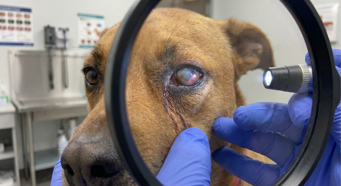
Introduction: The Threat of Corneal Ulcers
Corneal ulcers are a common and potentially devastating ocular condition in pets, ranging from superficial abrasions to deep, sight-threatening lesions. The cornea, the transparent outer layer of the eye, plays a crucial role in vision. Damage to this delicate structure can lead to severe pain, infection, and, if left untreated, irreversible blindness. While many ulcers respond well to medical management, there are critical instances where surgical intervention becomes not just an option, but an absolute necessity for saving the eye and preserving vision. This article delves into the various management strategies, highlighting the pivotal moments when surgery, from relatively non-invasive to complex reconstructive procedures, is vital for a successful outcome.
Understanding Corneal Ulcers in Pets
A corneal ulcer is essentially an open sore on the cornea, resulting from a loss of corneal tissue. These lesions are classified by their depth (superficial, stromal, descemetocele) and their nature (simple, complicated, indolent, melting).
Causes and Risk Factors
Common causes include trauma (e.g., scratches, foreign bodies), infections (bacterial, fungal, viral), eyelid abnormalities (e.g., entropion, distichiasis), tear film deficiencies (e.g., Keratoconjunctivitis Sicca or 'dry eye'), and underlying systemic diseases. Brachycephalic breeds (e.g., Pugs, Bulldogs, Shih Tzus) are particularly predisposed due to their prominent eyes and inability to fully blink, leaving their corneas vulnerable.
Initial Medical Management: The First Line of Defense
For superficial, uncomplicated corneal ulcers, medical management is typically the first line of defense. The primary goals are to prevent infection, control pain, and promote healing.
Standard Medical Therapies
This usually involves a combination of topical medications:
- Topical Antibiotics: To combat or prevent bacterial infections (e.g., neomycin, polymyxin B, tobramycin, ciprofloxacin).
- Pain Management: Topical atropine can reduce ciliary muscle spasm and pupil constriction, alleviating pain, especially in deeper ulcers. Oral NSAIDs may also be prescribed.
- Anti-collagenase Agents: For 'melting' ulcers (rapid breakdown of corneal stroma), medications like N-acetylcysteine or autologous serum/plasma are used to inhibit destructive enzymes.
- E-collar: Essential to prevent self-trauma and rubbing of the eye, which can worsen the ulcer.
Regular re-examinations are crucial to monitor progress and adjust treatment as needed.
When Medical Management Isn't Enough: Recognizing the Need for Surgery
While many ulcers heal within 7-10 days with appropriate medical therapy, others fail to respond or rapidly progress. These 'complicated' ulcers often necessitate a shift in strategy.
Indications for Surgical Intervention
Surgery becomes critical in several scenarios:
- Deep Stromal Ulcers: Ulcers extending into the deeper layers of the cornea.
- Melting Ulcers: Rapidly progressing ulcers characterized by enzymatic degradation of corneal tissue, posing an imminent threat of perforation.
- Descemetocele: An ulcer so deep that it has perforated all layers of the cornea except the innermost Descemet's membrane. This is an ophthalmic emergency, as rupture is highly probable.
- Perforated Ulcers: Full-thickness defects in the cornea, allowing aqueous humor to leak out and potentially leading to iris prolapse, endophthalmitis, and permanent vision loss.
- Non-healing or Indolent Ulcers: Superficial ulcers (often called Spontaneous Chronic Corneal Epithelial Defects - SCCEDs) that fail to heal after several weeks of appropriate medical therapy and conservative debridement.
In these cases, timely surgical intervention can prevent irreversible damage, salvage the eye, and restore comfort.
Critical Surgical Procedures for Advanced Ulcers
Conjunctival Pedicle Grafts (CPG)
One of the most common and versatile surgical techniques for deep or melting ulcers, and descemetoceles. A small piece of conjunctiva (the pink tissue lining the eyelids) is dissected, leaving one end attached to its blood supply, and then sutured over the corneal defect. The graft provides immediate structural support, brings a rich blood supply for healing, and delivers inflammatory cells and antibodies to fight infection. It is a highly effective method for preserving globe integrity and promoting rapid healing. Once healed, the pedicle graft can be partially or fully trimmed months later for aesthetic reasons, though some corneal scarring may remain.
Superficial Keratectomy
This procedure is primarily used for non-healing, indolent ulcers (SCCEDs) or for removing deep corneal foreign bodies, scar tissue, or pigmentary keratitis. It involves carefully debriding or excising the abnormal superficial corneal epithelium and a thin layer of underlying stroma. The goal is to remove the diseased tissue and provide a healthy bed for new epithelial cells to attach, promoting proper healing.
Corneal Transplants and Advanced Reconstructive Surgeries
For severe perforations, large defects, or cases where a conjunctival graft is insufficient, more advanced techniques may be necessary:
- Corneal-Conjunctival Transposition: Similar to a CPG but involves a larger, usually full-thickness, section of limbal conjunctiva and sclera, which can be rotated to cover a larger defect.
- Corneal Graft (Keratoplasty): Involves transplanting donor corneal tissue (from an eye bank) into the recipient's eye. This is a highly specialized procedure usually reserved for severe cases, offering the best potential for maintaining corneal transparency and vision following large full-thickness defects.
- Amniotic Membrane Grafts: Amniotic membrane can be used as a biological bandage to promote healing and reduce inflammation in some cases, often in conjunction with other procedures.
These procedures require meticulous surgical skill and specialized equipment, often performed by a veterinary ophthalmologist.
Post-Surgical Care and Long-term Management
Following surgery, intensive post-operative care is paramount for successful healing. This typically involves a regimen of topical antibiotics, anti-inflammatory medications, pain relief, and possibly atropine. An E-collar is absolutely mandatory to prevent self-trauma. Regular follow-up examinations are crucial to monitor the healing process, detect complications early, and ensure the best possible outcome.
Conclusion: Prioritizing Your Pet's Vision
Corneal ulcers in pets represent a spectrum of conditions, from minor abrasions to profound sight-threatening emergencies. While many can be managed medically, recognizing the critical signs that necessitate surgical intervention is key to preserving vision and ensuring your pet's comfort and quality of life. Deep, melting, descemetocele, perforated, and non-healing ulcers demand prompt and often surgical management by an experienced veterinary ophthalmologist. Early diagnosis, appropriate treatment, and diligent post-operative care are all vital components in navigating these challenging cases and giving your beloved companion the best chance for a healthy, pain-free eye.